
SQSP Example 3
/One of the greatest feats (and mysteries) of the human visual system is how we perceive a visually stable world despite constantly moving our eyes. Because we have the greatest visual acuity at our foveas, we repeatedly shift our eyes from location to location in order to have light from all aspects of our visual scene to land on them, allowing us to fully appreciate the visual aesthetic of, let’s say, the Golden Gate Bridge. Every eye movement, or saccade, results in a seamless and instant perception of the target visual area. Thus, despite a constant train of saccades, the locations of the cables and towers, the length of the bridge, and positions of the perched seagulls, all seem to maintain their relative sizes and distances from each other. The result is that we perceive the world as visually stable. Or do we? Recent work from the lab of Dr. Tirin Moore at Stanford University suggests otherwise.
The mechanisms that control eye movement are known to interact profoundly with those that control vision. Electrophysiological and psychophysical experiments involving awake, behaving macaque monkeys have played a key role in elucidating the ways eye movements modulate visual perception. In particular, the Frontal Eye Field (FEF), located in the arcuate sulcus of prefrontal cortex, is an area that has been shown to play a large role in mediating the cross talk between oculomotor and visual areas of the brain. The FEF contains a heterogeneous population of neurons, some which encode eye movement, also known as saccade, activity, others that encode visual activity, and others that encode both. It is the precise interaction between this unique pool of cells and various other visual areas throughout the brain that is thought to underlie many aspects of visual perception including visual attention, spatial working memory, exploration, and visual decision-making.
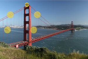
Because a subset of FEF neurons responds to visual stimuli, they, like neurons of visual cortex, have receptive fields (RFs). RFs are areas of the visual field in which neurons respond to particular visual stimuli. The retina maps visual space based on incoming light from the visual scene, and downstream visual neurons can integrate this information to encode specific subsets of the visual scene. Thus, RFs can be small or large, simple (e.g. vertical line) or complex (e.g. face). Importantly, this means that a neuron’s RF is defined by its inputs, and when the synaptic weights of these inputs change, the RF itself can change. When you combine all visual cells and their distinct RFs, the entire visual scene can be represented (Fig. 1). The question is, how is this visual representation maintained throughout a saccade?
As we move our eyes to a new location, we do not experience the displacement of the visual world during the shift from point A to point B. It is a perceptually instantaneous shift. It is widely assumed that a major mechanism underlying this effect is that the initial motor commands to our eyes are also sent to visual areas (a concept called corollary discharge) and cause a proactive shift of RFs to their new locations before the eyes actually move.
Thus, by the time we are actually executing a saccade, a visual neuron’s RF has already shifted to its new target location (Fig. 2).

This proactive shifting, termed predictive remapping, happens in less than a tenth of a second before you move your eyes! The idea is that without predictive remapping, the image of the Golden Gate Bridge would shear and distort as our eyes move from one location to the next because RF updating and visual processing would lag behind the actual eye position updating.
A recent study from the Moore lab, headed by postdoctoral fellow Marc Zirnsak, made a surprising discovery about visual FEF neurons that refutes traditional predictive remapping. In predictive remapping, RFs would presaccadically shift following a trajectory that is more or less parallel to and of the same magnitude as the saccade vector (Fig. 2). However, Zirnsak recorded temporally precise measurements of visual FEF neurons while monkey subjects executed saccades and showed that rather than RFs presaccadically shifting directly to their new retinocentric locations, they shift toward the saccade target location (Fig 3).
Zooming out to a larger scale and considering all visual FEF neurons, Zirnsak’s findings suggest that FEF RFs presaccadically converge onto the location of saccadic targets before settling into their new locations. The result: a brief, presaccadic compression of visual space representations in the FEF. For a split second before you move your eyes to a new location, the entire visual world is unnoticeably collapsing and compressing to that location (Fig. 4). Zirnsak et. al. hypothesize that this mechanism could facilitate a brief enhancement of visual processing at saccade target locations (which are areas of visual interest since we are saccading to them). A fascinating implication of these experiments is that this presaccadic compression of visual space may underlie a known perceptual correlate. Psychophysical experiments of the late 20th century showed that human subjects, tasked with pointing to the location of stimuli flashed briefly before saccades, localized the stimuli as being much closer to the saccade targets than they actually were. It is possible that the pressacadic compression observed in FEF is behind this. Thus, Zirnsak et. al. may have found a neural correlate underlying a psychophysical phenomenon that has been around for decades. In any case, these experiments have certainly changed the way we look at looking.
A recent study from the Moore lab, headed by postdoctoral fellow Marc Zirnsak, made a surprising discovery about visual FEF neurons that refutes traditional predictive remapping. In predictive remapping, RFs would presaccadically shift following a trajectory that is more or less parallel to and of the same magnitude as the saccade vector (Fig. 2). However, Zirnsak recorded temporally precise measurements of visual FEF neurons while monkey subjects executed saccades and showed that rather than RFs presaccadically shifting directly to their new retinocentric locations, they shift toward the saccade target location (Fig 3).


