Inflammatory comments on ageing

Cell-signalling.png

None of us is getting any younger. In fact, most of us can expect to live longer than ever. The world’s oldest man died just last week aged 116 and, as global life expectancy continues to increase, we’re going to have to tackle age-related frailty and functional fragility to avoid unbearable healthcare costs and provide quality of life for a growing population of centenarians. With rates of dementia as high as 60% in the over 85s1, neurodegeneration is a big issue; but treating age-related diseases in isolation may not be the most effective solution. Scientists now think of ageing as an intricate process during which tissues and cells change the way they communicate and interact, resulting in a gradual loss of function. Not exactly a simple cause-effect pairing that can be hit with a youth pill.  

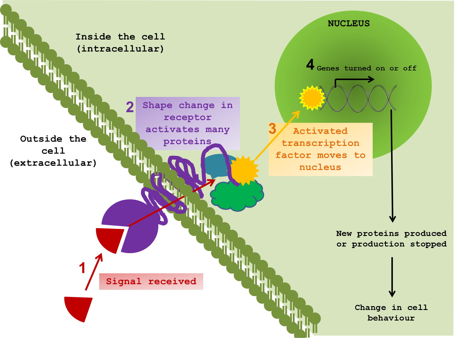
So how can we even start to pick apart the elaborate tapestry of ageing? What exactly happens when a cell changes the way it communicates? To perform effectively, neurons need to pick up signals from the outside world. Indeed all cells, tissues and organs function by responding to external signals, thus allowing an organism to respond and adapt to changes in environment. It is difficult to show in a static diagram the dynamic nature of these interactions. But I tried anyway. ↓

 Cell signalling

The majority of signals are chemical compounds, floating around outside a cell, waiting to contact their receptor protein. Once in range, the signal and the receptor are drawn closer and dock together. In response to the signal, the receptor twists and changes; not only on the surface, but also through the cell membrane and into the cytoplasm – the inner world of the cell. The shifting and gyrating receptor can hit other proteins, slapping on new chemical groups, removing others and sending these messengers on through the internal cellular space on a collision course with target proteins of their own. This molecular line of dominoes almost always ends at the cell’s control centre – the nucleus. It is here that messages are converted into changes in gene expression, protein production and, in turn, cellular behaviour. The proteins that directly interact with DNA to bring about such changes are called transcription factors.


Different signal molecules and receptors trigger distinct domino effects in the cell, which scientists refer to as signalling pathways. Though different cells are tuned to pick up different signals, many of the intracellular proteins used to transmit the message have been the same for millions of years. Throughout evolution, cells have taken the Heath Robinson* approach to signalling. Rather than inventing new proteins every time a new function had to be performed, cells used the tools already at their disposal. As a result, the same proteins can be found in different types of cells, across species and even across kingdoms. You share molecular signalling pathways with almost everything – from bears, to bananas to bacteria.

*Americans see Rube Goldberg


One versatile and highly abundant transcription factor, and the focus of a new study in Nature2, is NFkB (nuclear factor kappa B). This molecule is a hallmark of inflammation and once activated, perpetuates inflammatory responses. However, many different cells use this molecule to transmit all sorts of messages and, as a consequence, its role in a particular signalling pathway is often difficult to pin down. In their new study, Zhang and colleagues used several tricks to see just what NFkB is up to in the brain and how its activity relates to ageing. By linking it to a fluorescent molecule, the authors were able to visualise NFkB activity in the brain. The more active the transcription factor, the brighter the signal. In this case, brains got brighter with age supporting the idea that inflammation in the brain increases as we get older. While not always bad, inflammation in the brain is a sign of altered equilibrium and contributes to several neurodegenerative diseases3-6. By injecting a non-functional version of the protein, the authors were able to interrupt NFkB activation in the brains of old mice. Interestingly, reducing NFkB activity not only reduced inflammation, but also improved cognitive function (measured by maze navigation), decreased muscle degeneration (measured by grip tests) and even extended life span.


So why is inflammation increased in older brains? It turns out that in mouse middle age, the majority of NFkB activity comes from the microglia –the brain’s resident immune cells. A change in the calibration of these cells over time increases their sensitivity in old age. Instead of waiting for an infection, the microglia are active all the time and mount an inappropriate inflammatory response. This is when NFkB first gets activated and it makes the microglia produce other inflammatory molecules, which perpetuate the rumour that there is some kind of infection and that inflammation is necessary. Neurons take up these signals from the microglia and NFkB inside these cells also becomes active. This form of molecular baton passing between the immune microglia and the neurons of the hypothalamus is possible because they share the same signalling molecules, and is likely to bounce back and forth in an escalating feedback loop. In this way, an inappropriate response by one cell type (the microglia) can result in another cell type getting the wrong idea. As the signals feedback, the neurons change their behaviour. One consequence is that they gradually reduce production of an important molecule called gonadotropin-releasing hormone (GnRH). The decline of GnRH is responsible (along with other things) for the loss of reproductive function with age; but this new study showed that GnRH injection can also improve neural regeneration in old mice and strengthen cognitive and muscle function. In addition, by specifically removing a gene controlling NFkB in microglia, the authors were able to restrain inflammatory microglia in middle-aged mice. This nipped inflammation in the bud, prevented the involvement of neurons in the inflammatory process, reduced GnRH decline and extended lifespan. Hurray! A new miracle drug, right? Well, maybe not quite yet.


Unfortunately, we’re still a long way from using NFkB inhibition or GnRH replacement to extend human lifespan. The fact that many cells are using the same molecules to transmit messages makes targeting specific molecules very difficult. During evolution, different cell types adopted different roles as they became more specialised. However, having come from the same ancestral cell, they use the same old machinery to perform new and different tasks. Now, when a signal from one type of cell gets picked up by another, there is a level of cross-communication that can have unexpected and sometimes undesirable outcomes. If we try and remove a protein that is misbehaving in one cell type, we risk interrupting its routine function in another. The complex crosstalk between biological molecules is a caveat worth remembering whenever a new protein or gene hits the headlines as the root of all ageing or instigator of disease. These genes and proteins did not evolve to make us old and sick. They evolved to perform a task that is almost certainly essential for survival. It is the misuse of a protein or its appearance in the wrong cell at the wrong time that causes problems. A “disease-causing” protein is, in all probability, performing an essential role elsewhere in the body so cannot just be eliminated.


It will take incredibly sophisticated techniques to iron out exactly how best to recalibrate cellular function following an undesirable change like the increase in NFkB activity in older neurons. Unravelling the mysteries of ageing is not an easy task and is likely to involve the integration of many different scientific disciplines. By identifying how different cells and organs communicate, we hope to get a better idea of when and how signalling wires get crossed and so identify the right target to prevent disease and maybe even slow the ageing process.


1)         Alzheimer’s Association (2013). 2012 Alzheimer’s Disease Facts and Figures. http://www.alz.org/downloads/facts_figures_2012.pdf OPEN ACCESS!

2)         Zhang et al. (2013). Hypothalamic programming of systemic ageing involving IKK-β, NF-κB and GnRH. Nature 497: 211-216.

3)         Wright et al. (2013) Neuroinflammation and Neuronal Loss Precede Aβ Plaque Deposition in the hAPP-J20 Mouse Model of Alzheimer’s Disease. PLoS ONE 8(4): e59586. http://www.plosone.org/article/info%3Adoi%2F10.1371%2Fjournal.pone.0059586 OPEN ACCESS!

4)             Ugur Tufekci et al. (2012). Chapter Four – Inflammation in Parkinson's Disease. Advances in Protein Chemistry and Structural Biology Vol. 88 pp. 69–132. http://dx.doi.org/10.1016/B978-0-12-398314-5.00004-0

5)         Holmes (2013). Systemic inflammation and Alzheimer's disease. Neuropathology and Applied Neurobiology 39 (1): 1365-2990 http://dx.doi.org/10.1111/j.1365-2990.2012.01307.x

6)             Hauser and Oksenberg (2006). The Neurobiology of Multiple Sclerosis: Genes, Inflammation, and Neurodegeneration. Neuron 52 (1): 61–76 http://dx.doi.org/10.1016/j.neuron.2006.09.011

Brains & Bourbon Ep. 1: Attention and an Old Fashioned

Brains & Bourbon Ep. 1: Attention and an Old Fashioned

This summer we are launching a new podcast centered around the Stanford neuroscience community called "Brains and Bourbon," a show about cocktails and neuroscience. Each week, we invite a neuroscientist to discuss the process and motivation behind their science, and to share their favorite cocktail with us. Our first guest is Nick Steinmetz, a 6th year graduate student in Kwabena Boahen and Tirin Moore’s labs here at Stanford, who talks to us about attention and monkeys, and shows us how to make an Old Fashioned cocktail.

Read More

Linky and the Brain: Science and Art edition

Hi folks. Please excuse the brevity of this post - the adrenaline/endorphine one-two punch of tonight's softball game victory has worn off, and I'm ready to catch up on my sleep. But, before I pop off, here's some links I've been collecting over the past couple of weeks to share with you all.

Science? Art? Yes.

Via Laughing Squid, visualizations of animal sounds. The work of software designer and artist Mark Fischer, these eerily evocative illustrations are generated by passing animal calls through a wavelet analysis. Part work of art, part complex data visualization, I thoroughly enjoyed viewing these images. My particular favorite is the Blue-Crowned Manakin. Visit Aguasonic Acoustics for additional interpretations.

Again, courtesy of Laughing Squid, entries in Princeton University's 2013 Art of Science competition. 44 images selected from 170 submissions from 24 Princeton University departments, images were produced as part of scientific research. I vaguely remember Stanford University hosting such a competition; can someone more knowledgeable comment in some information? Shout out to my c.elegans homies (Hi, Sammy): I loved this picture of a swarm of worms on an agar plate. Also, here's a picture entitled "Worm Water Slide". Best title, or best title ever?

Over at the Smithsonian, a brief news report on research into how the natural frequency at which an individuals skull vibrates (this varies from person to person, from 35 to 65 times per second), can (moderately) predict what type of music the person does not enjoy listening to. The Unique Vibrations of Your Skull Affect How Your Hear Music.

Science, in the trenches.

I enjoyed reading this blog post, on the site Small Pond Science, that talks about the familiar situation of having a set of data that perfectly answers a hypothesis... that you didn't initially set out to test. Pretending you planned to test that hypothesis the whole time.

Slight/moderate facepalm moment: Cornell researcher and blogger Zhana Vrangalova writes up a post-game analysis of the media coverage of her recent paper, "Birds of a feather? Not when it comes to sexual permissiveness".

For pure delight, nothing can beat this, via The Atlantic: the second author of the recent Nature paper describing the collection of fluid isolated in the Earth's crust in the Precambrian era, "took one for the team" and drank some of their ~1 billion year old water. It doesn't taste very good.

Public and Private Sectors

A write up of a recent NSF-funded workshop on priorities for the Brain Initiative. (via nsf.gov)

Mozilla launches a new online resource for scientists, Science Lab, with the initial vision of encouraging researchers and members of the open web community to "share ideas, tools and best practices for using next-generation web solutions to solve real problems in science, and explore ways to make research faster, more agile and collaborative". (Press release, via The Next Web)

And that's all I've got. Until next time!


Comment

Astra Bryant

Astra Bryant is a graduate of the Stanford Neuroscience PhD program in the labs of Drs. Eric Knudsen and John Huguenard. She used in vitro slice electrophysiology to study the cellular and synaptic mechanisms linking cholinergic signaling and gamma oscillations – two processes critical for the control of gaze and attention, which are disrupted in many psychiatric disorders. She is a senior editor and the webmaster of the NeuWrite West Neuroblog

A Tale of Two Papers

A Tale of Two Papers

As behavioral neuroscientists, we hope that our findings generalize beyond the exact conditions of our experiments, and in many cases, beyond the species we choose to study. This is particularly true in labs that study models of psychiatric disease. Recent high profile co-publications on compulsive behavior and on depression, however, call this idea into question. Here I'll discuss these two pairs-of-papers, with an eye toward their implications for generalizability.

Read More

Ask Stanford Medicine: Dr. Josef Parvizi discusses drug-resistant epilepsy

As part of the Stanford School of Medicine's Ask Stanford Med series, Stanford neurologist Dr. Joseph Parvizi will be responding to questions on the subject of drug resistant epilepsy, and a surgical procedure to help treat the devastating condition. Dr. Parvizi, MD, PhD, is the director of the Stanford Program for Drug-Resistant Epilepsies. Dr. Parvizi will be taking questions both via twitter, or via the School of Medicine's Scope blog. Questions will be collected until Wednesday (June 19) at 5 PM Pacific Time. Both neuroscientists and members of the general public are encouraged to ask a question, and to join the conversation at the Scope blog.

To find out more about the event, or to ask a question or to join the conversation, visit the Scope blog.

To ask a question via Twitter, be sure to include #AskSUMed in your tweet.

Comment

Astra Bryant

Astra Bryant is a graduate of the Stanford Neuroscience PhD program in the labs of Drs. Eric Knudsen and John Huguenard. She used in vitro slice electrophysiology to study the cellular and synaptic mechanisms linking cholinergic signaling and gamma oscillations – two processes critical for the control of gaze and attention, which are disrupted in many psychiatric disorders. She is a senior editor and the webmaster of the NeuWrite West Neuroblog

The Cruel Irony of Saxitoxin

saxiotoxinblog.png

It was summer of 1997 at Cape Blanc on the western coast of Africa and some two hundred monk seals, nearly two-thirds of the population in the region, lay dead (1, 2). The cause of death was saxitoxin, a highly potent neurotoxin, whose existence is one of Nature’s cruelest ironies. Saxitoxin is a chemical compound, an alkaloid to be precise, that binds to sodium and potassium channels in the brain and muscle (3). This binding obstructs the flow of sodium and potassium ions across the cell membrane, impedes the ability of brain cells and muscle cells to send electrical signals, and can cause death by respiratory failure (3). Certain species of marine invertebrates and fish can accumulate saxitoxin to high levels and then pass it on to their predators. A possible mechanism for this is a difference in the sodium and potassium channels between predators and prey. Interestingly, some species of clam have a mutation in a single amino acid of their sodium channels that makes them resistant to saxitoxin (4), to the detriment of many animals up the food chain, including monk seals and humans. Ecologists estimate that during the fateful summer of 1997, the Cape Blanc monk seals could have been ingesting nearly lethal doses of saxitoxin every day from the fish they consumed (2). And every year, seafood contaminated with saxitoxin kills nearly 300 people and sickens another 1700 (3).

Where does saxitoxin come from? The villains in this story are marine microorganims called dinoflagellates. These microorganisms belong to the kingdom Protista, the dusty attic of taxonomy where biologists place eukaryotic life forms that they really don’t understand. Dinoflagellates are about as closely related to humans as to plants and fungi and incredibly diverse within themselves. About 4000 species have been described, 90% of which live in the world’s oceans and survive either by preying on other single-celled organisms or by doing photosynthesis (5). Most of these species do not produce saxitoxin but do have other curious characteristics, like genomes up to 100 times larger than the human genome, permanently condensed chromosomes in all stages of the life cycle (instead of just during cell division, like in other eukaryotes), and chloroplasts acquired independently of land plants (6). Three genera of dinoflagellates that do produce saxitoxin, Gymnodinium, Pyrodinium, and Alexandrium, get consumed by filter-feeding fish and marine invertebrates that effectively concentrate the poison in their viscera (2, 3). The monk seals at Cape Blanc had the misfortune to be near a bloom of Gymnodinium catenatum and possibly Pyrodinium bahamense (2). Such blooms occur seasonally and may account for other unexplained mass mortalities of marine wildlife (2), though, luckily for humans, many countries have monitoring programs for edible mussels, clams, gastropods, crustaceans, and fish that minimize public health risk (3).

Why do dinoflagellates produce saxitoxin? It is unlikely that they intend to kill marine mammals because the saxitoxin-producing species are photosynthetic (7). Arguably, they would derive no benefit from killing high-level predators in their ecosystems and may even be harmed by an increase in lower-level predators that would result. The large size of dinoflagellate genomes, the difficulty of culturing dinoflagellates in the lab, and their extreme divergence from all organisms with sequenced genomes makes genomic and evolutionary studies of saxitoxin production in dinoflagellates challenging, and information is incomplete (8). The most popular and widely studied hypothesis is that saxitoxin deters the immediate predators of photosynthetic dinoflagellates, such as cocepods and mollusks (3). However, evidence is inconclusive because though saxitoxin production correlates with decreased consumption of dinoflagellates by cocepods, many studies have found no effect of saxitoxin on the survival of mollusks, and other studies found that even dinoflagellate species that do not produce saxitoxin could kill their predators by an as-yet-unidentified mechanism (3). Another hypothesis is that saxitoxin acts as a pheromone and regulates mating and other social behavior in dinoflagellate colonies, though any hypothesis that proposes a non-toxic role for saxitoxin must account for how dinoflagellates that do not produce it perform the same tasks (3). Nonetheless, at present, it is a good bet that dinoflagellates use saxitoxin for their own purposes and that its effect on the brain is a cruel irony.

 Sources

  1. The IUCN Red List of Threatened Species. http://www.iucnredlist.org/details/13653/0
  2. Reyero M et al (1999). Evidence of Saxitoxin Derivatives as Causative Agents in the 1997 Mass Mortality of Monk Seals in the Cape Blanc Peninsula. Natural Toxins. 7: 311-315. Open access.
  3. Cusick KD and GS Sayler (2013). An Overview on the Marine Neurotoxin, Saxitoxin: Genetics, Molecular Targets, Methods of Detection and Ecological Functions. Marine Drugs. 11: 991-1018. Open access.
  4. Bricelj VM et al (2005). Sodium channel mutation leading to saxitoxin resistance in clams increases risk of PSP. Nature 434:763-767. Paywall.
  5. Dinoflagellates. Smithsonian Museum of Natural History. http://www.mnh.si.edu/highlight/sem/dinoflagellates.html
  6. Wisecaver JH and JD Hackett (2013). Dinoflagellate Genome Evolution. Annual review of microbiology. 65: 369-387. Paywall.
  7. Algae and Human Affairs edited by CA Lembi and JR Waaland and published in 1989.
  8. Hackett JD et al (2013). Evolution of saxitoxin synthesis in cyanobacteria and dinoflagellates. Molecular biology and evolution. 30: 70-78. Open access.

Olds & Milner, 1954: “reward centers” in the brain and lessons for modern neuroscience

Sometimes the discoveries most exciting to read about are those that were made long ago, due to the sheer advance in knowledge that they represented. Such classic studies also remind us that the most important discoveries can be made with even the most rudimentary techniques, when combined with careful observation and clever interpretation. In this post, I will summarize and provide brief commentary on a classic paper in the field of neuroscience by Olds and Milner in 1954.

Read More

Tiger tiger, burning bright: the retinal specializations that cause glowing eyes

Tiger tiger, burning bright: the retinal specializations that cause glowing eyes

Bobcats like to chase laser pointers. At least, Moses the bobcat, of Big Cat Rescue, does. As entertaining as I found the sight of a bobcat pouncing at a glowing red dot (which is to say, very), I was more struck by the sight of the eyes, luminous in the darkness. Like any proper biologist, I googled “feline eye glowing”. Allow me to introduce the tapetum lucidum a layer of highly reflective tissue found behind the retina of many vertebrate species that provides, essentially, a second chance at photon detection.

Read More

Ask a Neuroscientist! – Seeing Colors

Ask a Neuroscientist!  –  Seeing Colors

Ask a neuroscientist! is a new column where we answer your questions about neuroscience! Our first question comes from personal correspondence from Michael in Azusa, California. Michael asks: “I recently read a news article about a woman who could see more colors of the spectrum than a normal person can. Can scientists see a difference in the brains of someone who can see more colors compared to a normal person? What about someone who is colorblind?”

Read More