Linky and the Brain: a grab bag from drunk science to itchy bears
/
This week's pickings from around the web. As esoteric as you like it.
Read MoreHome of the Stanford Neuroblog. Scientists writing about science, for a general audience.
Have a question about the brain? Ask a neuroscientist a question.
This week's pickings from around the web. As esoteric as you like it.
Read More
I’m really only half joking when I say I want to be an astronaut when I grow up. When experiments aren’t going well, my friends and I discuss the various ways in which we could convince the International Space Station (ISS) that we’re Star Fleet material. Since the relaxation of military and flight time requirements, we’ve been looking forward to stepping off this chaos of hard clay to wander darkling in the eternal space. Romance aside, the effects of space travel on health and the unique physiological and psychological conditions of long-range space travel really do interest a growing number of research scientists, myself included. How exciting, then, to hear from my friend Rishi that CASIS (the Center for the Advancement of Science in Space) have issued a request for proposals looking into the effects of microgravity on stem cells. I’m a biologist; surely I work on stem cells. Well, not quite. I work on the immune system, the cells of which do indeed derive from stem cells, but my interests lie much further down the developmental line. I study how a fully mature immune system works to protect the body against infection and how vaccines use the same machinery to protect against diseases before we encounter them. To me, this has obvious applications for interplanetary space exploration.

A recent publication in the Journal of Clinical Immunology1 shows altered immune function following space flight. Levels of inflammation-driving molecules in the blood go up, but specific responses to viruses by T cells go down. Is this a Big Deal? Let’s assume that people aren’t allowed into space if they have a serious illness and that the vacuum of space is clean enough that disease-causing bugs are unlikely to get on board a spaceship and make everyone evolve backwards or suddenly challenge the entire crew to a duel. Does it matter if the immune system is depressed in space? Well, one of the observations highlighted in the paper is that virus-specific responses are reduced. This might not be a problem if we weren’t all riddled with a large number of viruses that are only kept in check by constant immune surveillance. Almost everyone is infected with JC virus, Cytomegalovirus, Epstein-Barr virus, Varicella-Zoster virus, and several other strains of common herpesviruses. Many diseases, including shingles, are associated with a drop in T cell responses and can be severely debilitating. Surely we need someone doing research up there to see how immune responses to these long-term resident viruses change. An outbreak of shingles during a long voyage may not make compelling television, but could severely compromise an astronaut’s ability to function when far from access to antiviral drugs and pain medication. Clearly (if perhaps tinged with a little bias), research into the immune system is much more directly applicable to survival in space than stem cell research, which is still very much in the early stages.
As Commander Chris Hadfield showed so wonderfully, there are many and varied experiments that can be conducted in space that will have an impact on future astronauts. Part of me is tempted to speculate as to why CASIS have limited their remit to stem cells. Is it just because they’re cool and in the news a lot these days? Have the people controlling the purse strings been sweet-talked by a lobbyist with stem cell leanings? Or was this really the result of a long and in-depth study section on the biological priorities of extraterrestrial research? The cynic in me is clamouring for a rant about the whims of science policy but, since I know so little about the process, I should probably save my energies for more productive tasks. Like thinking of ways to convince CASIS that I work on stem cells.
References
1) Crucian et al (2013). Immune system dyregulation occurs during short duration spaceflight on board the space shuttle. Journal of Clinical Immunology 33 (2): 456-465.
Editor's Note: The author wishes me to express that she would prefer that CASIS used the correct spelling of 'Centre' in its name. Unfortunately, there was not time for the Center to make this official name change prior to this article going to press. Watch this space for updates.
I want to let you in on a little secret. We neuroscientists are actually quite jealous of the physicists. They may lament the fact that their unified theory of everything hasn’t turned up yet, but they’re sitting pretty with a bevy of universal laws, forces, constants and equations that do a bang up job explaining the universe. We neuroscientists are still on the hunt for some all-encompassing laws and principles that would explain brain function at a larger scale than the operation of single neurons (on which, I must say, we’ve done a pretty awesome job).
Read More
Hi folks! The theme of my Linky and the Brain this week is: model organisms, a love/hate relationship.
First off, I will direct you to Kelly Zalocusky’s hot-off-the-presses Neuroblog post, Of Mice and Men: On the Validity of Animal Models of Psychiatric Disease. Kelly discusses the difference between homology and analogy, and how the distinction between the two may be at the heart of why the translation between model organism and human disease state is such a difficult one. My newest goal for the Neuroblog is to start getting some conversations going in the comments – can we all meet up at Kelly’s post and chat about evaluating our model systems in terms of homology/analogy? I, for one, think about this question often (read: whenever I’m writing a grant). I’d love to learn where other folks are on the axis of “strive to be as close to the human disease state as possible so as to increase chances of translation” versus “not-strictly homologous/translation-able is fine, as long as we are learning basic things about the brain that will reasonably contribute to our greater understanding of how brains work”.
On the subject of model organisms, and more accurately dissatisfaction with model organisms, I read, with great interest, an article in the New York Times about researchers who develop allergies during the course of their research, to model organisms or common lab substances. Now, this concept is hardly new to me, or likely to anyone who has ever worked in a rodent lab. (I work closely with a postdoc who is so allergic to rodents he wears Teflon-coated gloves when handling them, to prevent contact/scratches.) What I found so interesting, was that the New York Times published the article in the first place; a trend piece about the travails of the common researcher. Awesome.
To read about a researcher who became allergic to cicadas, plus other stories, check out Allergies in the Time of Research, by Hillary Rosner.
One more feature regarding model organisms, this time my own. From the blog Last Word on Nothing, a delightful post describing the glory that is The Art of Chicken Sexing. Go for a description of a process so mysterious, that even the practitioners themselves can’t describe what they are looking for. Stay for the quotes from folks for whom the process of chicken sexing has become either an addiction, or a compulsion (or both).
And lastly, a hysterically funny post by scicurious, over at Scientopia, entitled Mopey Mice Pee Their Feelings. A blog post that is all about a novel method for evaluating the anxiety/depression in rodents. The method: urine tracking. Some of my favorite quotes from the post:
To introduce the concept of rodent urination:
"If you've held a lot of mice, you've been peed on a lot. Everywhere you put a mouse, that mouse WILL pee. It's part of the game and one of the things you get used to (probably one of the things we should warn new grad students about, too. "Congratulations! Be prepared to be peed on!")."
To describe a figure of the urination pattern of a "control" mouse:
"… a mouse I would have nicknamed "the dragger". He doesn't sprinkle it around, he DRAGS it around, and he is letting that lady know that he is HERE and READY."
And finally:
"It's like mousey feelings on paper."

As biomedical researchers, we use animal models as a compromise. We hope to understand human disorders and improve human health, but the experiments we do are often too risky for human subjects. One largely unspoken concern about this compromise is the degree to which these animals’ behaviors accurately model the disorder in question. What do we even mean when we say that a particular rodent behavior “models” a human syndrome? And why is it that, very often, treatments that work in animal models fail once they reach the clinical setting (1)?
There is an extensive literature in psychology on the various ways to assess the validity of tests and models (2), and the biomedical research community would do well to consider this long philosophical struggle. But, as a behavioral ecologist and ethologist, there seems to be one potential gold-standard question for animal models that is rarely, if ever, discussed. Are apparent similarities between the human and the animal behavior driven by homology, or are they analogies, driven by convergent evolution?
As I see it, one major flaw in the design of animal models is in mistaking analogy for homology. That is, neuroscientists often study an animal’s behavior because it resembles an interesting human behavior. Take, for example, mouse models of obsessive-compulsive disorder. The goal is not to understand why some mice groom too much, but instead to understand why some humans wash their hands too much. Mouse grooming is an analogy for hand washing. These studies are only useful, then, if mouse grooming and human hand-washing rely on the same neural circuitry. For these studies to be meaningful, the two behaviors must be homologous.
What does it mean to be homologous?

Homology means evolved from the same ancestral structure or behavior. If, for example, you wanted to understand the structure of bat wings, but could not get the permits to study bats, you could reasonably study bird wings as a model. You could also study human arms, or even whale flippers. The only reason such studies would be useful is that bat wings, bird wings, human arms, and whale flippers have very similar, evolutionarily homologous, structures (see figure). Even though whale flippers are not used for flight (“And the rest, after a sudden wet thud, was silence…”), their structure can tell you a lot about how bat wings are likely put together.
An analogous behavior or structure, on the other hand, is one that looks similar across species but likely occurs for different reasons or through entirely different mechanisms. A bat wing and a butterfly wing are analogous—while they look similar, and evolved to promote the same behavior, they are evolutionarily and structurally distinct. Attempting to learn about the skeletal structure of bats’ wings by studying butterflies would be a largely fruitless endeavor.
The difficulty, of course, in studying psychiatric disease is that most psychiatric diseases are defined by a cluster of symptoms—not by an underlying physiological process. For the researcher, this means that it is challenging to know whether you are studying the right physiological process at all. If a particular assay, based originally on analogy, repeatedly fails to translate in clinical trials—for example, if social behavior assays in mouse autism, or over-grooming in mouse OCD, or refusing to swim in mouse depression repeatedly let clinicians down—perhaps we, as a community, should consider this potential reason why.
Why would a drug designed to prevent and treat malaria, a parasitic infection of the blood and liver, also affect the central nervous system? The drug in question is mefloquine, marketed as Lariam, and I first learned about its bizarre side effects, including amnesia, psychosis, and hallucinations, while listening to “Contents Unknown,” an episode of the radio program This American Life. The episode intrigued me because it told the story of David MacLean, who was taking mefloquine while on a Fulbright scholarship in India and one day found himself in a train station in a different city from where he lived with no memory of who he was or how he got there. I went looking for primary scientific literature on how mefloquine affects both the malarial parasite and the human brain, and here is what I found. Before exploring the side effects of mefloquine, let’s tackle a more basic question: why do drugs have side effects? The answer lies in how drugs are discovered. In a perfect world, scientists would know so much about a disease that they would be able to design a precisely targeted drug, highly effective against the cause of the disease and benign for the patient. That is a major goal of biomedical research and highly desirable for malaria, which every year afflicts half a billion people (1) and kills one million children in Africa alone (2), but much more of this research is still needed. For many diseases, sadly including malaria, our knowledge is too limited to allow rational design of drugs, and most drugs are discovered either accidentally, like the first anti-malarial agent quinine (3), or by trial and error, which means taking some chemical compound, using it against the disease in an animal model, and seeing if the animal gets better. Therefore, a chemical compound often becomes a drug not because we understand how it works against the disease but rather because we have observed it to work. For a drug discovered in this way, we do not know whether its desired effect is its only effect until we try it out.
Mefloquine is an example of the trial-and-error approach. In the 1960s and 1970s, the Walter Reed Army Institute of Research tested over 300,000 chemical compounds for their ability to kill Plasmodium falciparum and Plasmodium vivax, the two most common malarial parasites, in owl monkeys, the best animal model at the time (4). Mefloquine showed the most promise and went on to clinical trials in humans that are meant, among other things, to test for side effects. In the case of mefloquine, these initial clinical trials showed no serious side effects, but they were conducted in vulnerable populations unable to give full consent, namely male prisoners, military personnel, and residents of developing countries, and may have been biased (5). More recent epidemiological research has shown that side effects in the central nervous system severe enough to require hospitalization occur in 1:10,000 patients taking mefloquine for malaria prevention and in 1:200 to 1:1200 patients using mefloquine for malaria treatment (6). Though mefloquine is still widely available, medical practitioners have an increased appreciation of its side effects, and it is now the drug of last resort, rather than of choice, for the U.S. military (5).
How does mefloquine act to kill Plasmodium parasites? This turned out to be a hard question to answer. Mefloquine is thought to inhibit growth of Plasmodium inside human red blood cells (7). The Plasmodium parasites have a complicated life cycle, proceeding from the saliva of mosquitoes to penetrate inside human red blood cells and liver cells. One possible target of mefloquine may be the food vacuole, a sort of microscopic stomach inside Plasmodium cells where they digest nutrients obtained from the cytoplasm of red blood cells, because the food vacuole changes shape in response to mefloquine treatment (8). I was unable to find any publications that identified the targets of mefloquine more specifically. This may be because laboratory experiments on Plasmodium species are difficult. My classmate Hao Li, a graduate student in the lab of Professor Matt Bogyo at Stanford, works with Plasmodium falciparum and has often described that the procedure of culturing it in blood cells is laborious, yielding precious little material for experimentation. And that’s the easiest parasite species to cultivate. The only way to obtain Plasmodium vivax for experiments is to let it infect and reproduce inside mice or monkeys (1).
Research on how mefloquine may be causing central nervous system side effects in humans was somewhat easier to find, though it is far from conclusive. Mefloquine doesn’t dissolve well in water but sticks quite well to the outside of blood cells and brain cells (5). Post-mortem examinations of both mice and humans exposed to mefloquine have found it to accumulate in the limbic system, a region of the brain responsible for emotions and memory (5) (for a bit more background on the limbic system, see my post “Linguistic Disconnect between the Brain and Emotions”). There it may be blocking connexins (5), proteins that are components of gap junctions. Gap junctions are channels in neuronal membranes that go from the cytoplasm of one neuron into the cytoplasm of the adjacent one. Thus, gap junctions are responsible for synchronizing the activity of neurons (5). Cap junctions are channels that form direct links between the cytoplasms of neighboring cells. Gap junctions are a critical pathway for direct cell-to-cell communication in both neurons (5) and glia (10). These channels pass both electrical current (in the form of charged ions) and intracellular signaling molecules, playing a role in synchronizing neuronal activity, as well as metabolic coupling and chemical signaling (11).
The blocking effect of mefloquine is strong enough and specific enough to have been used to study the signaling behavior of connexins (9). The blockage of connexins may impede the ability of the brain to control emotional impulses and may interfere with memory formation (5). However, the picture may be more complicated because I also found publications claiming blockage of a different class of proteins, called 5-HT3 receptors (7), and an effect on the ability of rat neurons to control their internal concentration of calcium ions, which is essential for neuronal signaling (6). Hopefully, future research will elucidate the relative importance of these various effects.
Current understanding of both the desired effect and the side effects of mefloquine is incomplete and more research is needed. But mefloquine also is a cautionary tale of pitfalls in the process of drug design that may misrepresent a compound with dangerous side effects as a perfectly safe one. It is an illustration of how much more we still need to understand about the human body and its parasites to be able to effectively treat malaria without driving anyone insane.
Hello all – back from a nuptial break! Here are some of the eye-catchers that have drifted across my screen recently:
Read MoreI’ve been thinking a lot about acronyms. It seems to me, that an acronym is a super sexy thing to have.
In the past few months, my little corner of Neuroscience has enjoyed the appearance of 4 new acronyms. Now, I’m not talking about the consonant-and-vowel salad used as shorthand for ever-more-sub-classified brain regions. Although, as a side note, I learned last Thursday during a lab meeting presentation that no matter how firm my intentions, I cannot seem to say dlPFC. Instead of a neat word-limit-approved acronym, I find myself pronouncing dorsolateral prefrontal cortex in a burst of desperate enunciation.
But the acronyms that float through my NCBI email alerts are not these brain region shorthands. Rather, they are the badges of newly minted techniques, or ideas, branded with a catchy, sexy acronym to help propel them into the neuroscience community’s awareness.
(Aside - Attention: D. Bochner.)
There are a lot of neuroscientists out there. And all of us are constantly producing new research, new techniques, new publications. Standing out amongst all this noise can be an imperative; critical for attaining an elusive funding source, or a prized (by some) academic job. So it makes perfect sense to me that a researcher, armed with a novel technique (or combination of ideas) would seek to use a clever acronym as a way to catch the attention of her/his fellow researchers. Can we together admit to a feeling of professional jealous admiration for a colleague armed with an acronym? (Catherine, nicely done with SPLURgE. Casey, TRAP is so delightfully customizable: ArcTRAP, fosTRAP. Excellent work.)
Plus, how thrilling to hear your colleagues utter the creative symbol of your research efforts. The next best thing, perhaps, to the nomenclatural heights enjoyed by Golgi, Nissl, Purkinje, Brodmann; that age of naming, it seems, has passed. (Although vestiges persist; see the newly discovered Dua’s layer, so found and named by Harminder Singh Dua, U. Nottingham).
Acronyms are also clearly useful for condensing a complex technique into an easily stated verbal handle that can be readily passed along to PR departments, or thrown around in scientific discussions. My personal struggles with dlPFC aside, I think we can all agree that including “clear lipid-exchanged acrylamide-hybridized rigid imaging/immunostaining/in situ hybridization-compatible tissue-hydrogel” in a sentence would be a trial. Far easier to say “CLARITY”. And with the added benefit of an evocative name to entice readers, prospective users, and public audiences. Here too we find the BRAIN Initiative (Brain Research through Advancing Innovative Neurotechnologies). Open to some good-natured ribbing for using “Brain” as the B in BRAIN? Sure. But ever so ready for prime-time publicity.
The other week, during a grant application planning session, I was counseled that using a particular acronym-ed technique (hopefully soon to be published from my lab), could help my grants score. An acronym bringing a sense of the official, the innovative, where a colloquial description of a technique is merely… there. After receiving Summary Statements that praise my scientific questions, only to declaim the innovation of my techniques, I can’t argue with the impulse. Will the technique be critically necessary to answer my scientific questions? Maybe. Nevertheless, as I construct my research strategy, I’ll be spending some quality time considering whether a sexy acronym could catch the attention of that NIH review committee, and secure me the funding I need to help my lab stay in business.
SPLURgE. From Chrisitan et al (2013). Sniffer Patch Laser Uncaging REsponse (SPLURge): an assay of regional differences in allosteric receptor modulation and neurotransmitter clearance. J Neurophysiol. Epub ahead of print. Link
TRAP. From Guenthner et al (2013). Permanent Genetic Access to Transiently Active Neurons via TRAP: Targeted Recombination in Active Populations. Neuron. 78(5):773-84. See previous blog post. Or go see the paper directly.
CLARITY. From Chung et al (2013). Structural and molecular interrogation of intact biological systems. Nature. 497(7449):332-7. Link
BRAIN Initiative. Via the NIH and the White House.
Part 7 in an occasional feature, highlighting recently published articles featuring an author (or authors) affiliated with the Stanford Neuroscience Ph.D program. This round, we've got two lovely first author papers, by Casey Guenthner and Izumi Toyoda.
Let's begin with my year-mate, Casey Guenthner (Luo Lab), who published his development of a technique, TRAP (Targeted Recombination in Active Populations), that allows genetic targeting of populations of neurons that are defined by whether they were activated in vivo by a set stimulus. For details, check out Casey's abstract, below.
Targeting genetically encoded tools for neural circuit dissection to relevant cellular populations is a major challenge in neurobiology. We developed an approach, targeted recombination in active populations (TRAP), to obtain genetic access to neurons that were activated by defined stimuli. This method utilizes mice in which the tamoxifen-dependent recombinase CreER(T2) is expressed in an activity-dependent manner from the loci of the immediate early genes Arc and Fos. Active cells that express CreER(T2) can only undergo recombination when tamoxifen is present, allowing genetic access to neurons that are active during a time window of less than 12 hr. We show that TRAP can provide selective access to neurons activated by specific somatosensory, visual, and auditory stimuli and by experience in a novel environment. When combined with tools for labeling, tracing, recording, and manipulating neurons, TRAP offers a powerful approach for understanding how the brain processes information and generates behavior.
I will leave you all with the comment that Casey's cartoon of the mouse whisker pad is glorious in its anatomical accuracy. And it's pretty cute, too. Figure 3b. Check it out below, along with data demonstrating targeting of active neurons in the whisker barrel system.

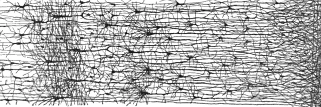
Just last week, Izumi Toyoda (Buckmaster lab) published her work recording spontaneous seizures in rats with temporal lobe epilepsy. Using 32 recording electrodes per rat, Izumi records from a massive number of brain structures, tracking the spread of seizures within the rodent brain. Her paper compares the propagation of the spontaneous seizures in the rats to known seizure activity in human patients with temporal lobe epilepsy. The results validate the pilocarpine model of temporal lobe epilepsy, showing seizures begin in similar brain locations in human patients and rodent subjects.
Temporal lobe epilepsy is the most common form of epilepsy in adults. The pilocarpine-treated rat model is used frequently to investigate temporal lobe epilepsy. The validity of the pilocarpine model has been challenged based largely on concerns that seizures might initiate in different brain regions in rats than in patients. The present study used 32 recording electrodes per rat to evaluate spontaneous seizures in various brain regions including the septum, dorsomedial thalamus, amygdala, olfactory cortex, dorsal and ventral hippocampus, substantia nigra, entorhinal cortex, and ventral subiculum. Compared with published results from patients, seizures in rats tended to be shorter, spread faster and more extensively, generate behavioral manifestations more quickly, and produce generalized convulsions more frequently. Similarities to patients included electrographic waveform patterns at seizure onset, variability in sites of earliest seizure activity within individuals, and variability in patterns of seizure spread. Like patients, the earliest seizure activity in rats was recorded most frequently within the hippocampal formation. The ventral hippocampus and ventral subiculum displayed the earliest seizure activity. Amygdala, olfactory cortex, and septum occasionally displayed early seizure latencies, but not above chance levels. Substantia nigra and dorsomedial thalamus demonstrated consistently late seizure onsets, suggesting their unlikely involvement in seizure initiation. The results of the present study reveal similarities in onset sites of spontaneous seizures in patients with temporal lobe epilepsy and pilocarpine-treated rats that support the model's validity.
And because I showed a figure from Casey's paper, here is one from Izumi's, highlighting a subset of 16 recording electrodes on which a spontaneous seizure is recorded in an epileptic, pilocarpine-treated rat. Let there be no doubt in your mind - the skill required to implant 32 recording electrodes into a rat is large. I suggest being very impressed with Izumi's surgical skills.

Dr. Caitlin O'Connell-Rodwell, professor of Otolaryngology at Stanford University, studies seismic communication in elephants.This post collects videos of Dr. O'Connell-Rodwell discussing her research, as well as a brief discussion of the basics of seismic communication in elephants.
Read MoreHome of NeuWrite West and the Stanford Neuroblog.
Do you have burning questions about how the brain works? You’ve come to the right place! Submit all your questions to NeuWrite West and we will have a neuroscientist research and answer your question.
Note: The NeuWrite West team consists of research scientists. Therefore, we can not provide answers to questions of a medical nature; we are not medical doctors.
Questions will have a turn-around time of approximately one month.
Or search for "NeuWriteWest" (without spaces) on your favorite podcast app!
Powered by Squarespace